
Oral Pathology
Expert Oral Pathology Care
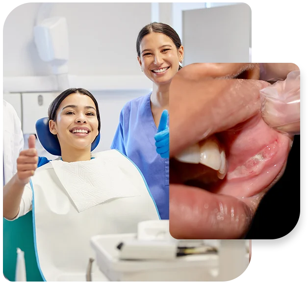
Oral Pathology for Soft and Hard Tissues
Dr. Patel manages a wide variety of pathology associated with the oral cavity. Patients are often referred by their dentist or physician for the management of soft or hard tissue abnormalities for biopsy. Soft tissue lesions generally involve ulcers, patches, bumps, and discoloration on the gums, lips, cheek, tongue or salivary glands. Hard tissue pathology encompasses alterations in teeth or bone. Abnormal changes in the mouth typically are detected early as the oral cavity is readily accessible. Oral biopsy in the mouth, soft palate, and throat can be carried out comfortably with local anesthesia, and with sedation if indicated. Usually, minimal recovery time is needed.
aid in early recognition
Tissue changes do not always mean cancer, but certainly need to be evaluated promptly to determine the cause. Since oral cancer kills one person every hour, twenty-four hours a day, in North America, Dr. Patel strongly encourages performing self-examinations monthly to aid in early recognition as well as routine annual office screening.
Precision and Expertise
Specialized Skills & Professional Care for Oral Surgery
Self-Examinations
- red or white patches of the oral tissues
- a sore that fails to heal and bleeds easily
- an abnormal lump or thickening of the tissues of the mouth
- chronic sore throat or hoarseness
- difficulty in chewing or swallowing
- a mass or lump in the neck